25th Annual Scientific Assembly
Ultrasound — Advanced
Sunday, March 10, 2019
8:00am-12:30pm
Forum 4-5
Note: Advanced registration is full for this pre-conference course.
Course Description
This year’s AAEM pre-conference ultrasound course has been fully updated to reflect participants’ wishes in designing the ultimate ultrasound course. Each year after reviewing participant comments we construct a new course to address their needs.
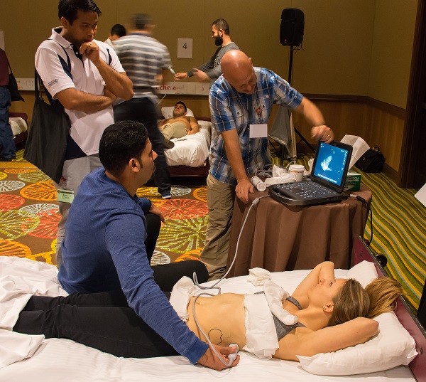
Participants loved last year’s course and we have added more modules. Didactic lectures will take place online at your convenience. The lectures will be available one month prior and one month following the advanced US course. There will be a maximum four participants to one instructor allowing each individual participant ample hands-on time for ultrasound scanning.
← Back to all pre-conference courses

Register
Note: The Ultrasound — Advanced Course is full at this time. To be added to the wait list, please email Zenita Hill at zhill@aaem.org. In your email, please include the five modules you are interested in.
Credit Designation Statement
The American Academy of Emergency Medicine (AAEM) designates this live activity for a maximum of 3.75 AMA PRA Category 1 Credit(s)™. Physicians should claim only the credit commensurate with the extent of their participation in the activity.
Course Fee
- AAEM and AAEM/RSA Members: Early Bird $375 | Late: (after February 6) $475
- Non-AAEM Member: Early Bird: $525 | Late: (after February 6) $625
- Non-AAEM/RSA Resident Member: Early Bird: $435 | Late: (after February 6) $535
- Non-AAEM/RSA Studnet Member: Early Bird: $415 | Late: (after February 6) $515
Special Discount if taking both Ultrasound — Beginner and Advanced
- AAEM Member: Early Bird $600 | Late: (after February 6) $700
- Non-AAEM Member: Early Bird: $750 | Late: (after February 6) $850
Tentative Course Schedule
Sunday, March 10, 2019
|
8:00am-8:15am
|
Welcome
Michael J. Lambert, MD RDMS FAAEM and Katharine Burns, MD FAAEM
|
|
8:15am-9:00am
|
Hands-on Module 1
|
|
9:00am-9:45am
|
Hands-on Module 2
|
|
9:45am-10:30am
|
Hands-on Module 3
|
|
10:30am-10:45am
|
Coffee Break
|
|
10:45am-11:30am
|
Hands-on Module 4
|
|
11:30am-12:15pm
|
Hands-on Module 5
|
|
12:15pm-12:30pm
|
Wrap up & Adjourn
Michael J. Lambert, MD RDMS FAAEM and Katharine Burns, MD FAAEM
|
Pick 5 application modules. Modules are selected in the online registration form.
- Aorta & IVC
- Cardiac-Advanced
- DVT
- eFast
- Gallbladder & Renal
- Gastrointestinal
- Head & Neck
- Image Acquisition & Instrumentation
- Landmark Documentation
- Musculoskeletal — General
- Musculoskeletal — Shoulder
- Ocular
- Procedures — Nerve Blocks
- Procedures — Non-Vascular
- Procedures — Vascular Access
- Pulmonary
- Shock
- Transesophageal Echocardiography (TEE)
Learning Objectives
Aorta & Inferior Vena Cava
- Understand the surface landmarks for appropriate transducer positioning to perform sonographic examinations of the abdominal aorta and Inferior vena cava.
- Demonstrate the ability to identify and visualize landmarks for the aorta and IVC in the transverse and longitudinal scanning planes.
- Understand the sonographic findings and pitfalls for identifying pathology including aortic aneurysm.
- Understand the utility of motion modality (M-mode) and demonstrate its use.
- Acquire and interpret sonographic images of heart (subcostal) and IVC in the transverse and longitudinal planes.
- Identify volume status of the IVC based on size and responsiveness to fluid.
Cardiac-Advanced
- Understand the utility of motion modality (M-mode) and demonstrate its use.
- Demonstrate the surface landmarks and transducer position necessary to perform an echocardiogram.
- Acquire and interpret sonographic images of heart (subcostal, parasternal long, parasternal short and apical windows).
- Identify pathologic conditions such as pericardial effusion, gross wall motion abnormalities and cardiac tamponade.
- Demonstrate landmarks and measurements for cardiac output.
- Understand US findings for diastolic and systolic heart failure.
DVT
- Understand the sonographic landmarks and anatomical relationships as they relate to the vasculature of the neck, upper extremity and lower extremity.
- Acquire and interpret sonographic images of the internal jugular, femoral, basilic, brachial and axillary veins in live patient models.
- Demonstrate compression technique of upper and lower extremity veins.
eFast
- Understand the surface landmarks for appropriate transducer positioning to perform the FAST examination.
- Understand the sonographic landmarks and anatomical relationships of the heart, liver, spleen and bladder as they relate to the FAST examination.
- Demonstrate the ability to identify and visualize the areas of potential intra-abdominal and thoracic spaces for free fluid to collect or pneumothorax.
- Understand the sonographic findings and pitfalls for identifying life-threatening trauma conditions such as cardiac tamponade, hemo/pneumothorax and intra-abdominal hemorrhage.
Gallbladder & Renal
- Understand the surface landmarks for appropriate transducer positioning to perform sonographic examinations of the aorta, kidney and gallbladder.
- Understand the sonographic windows and landmarks of the aorta, kidney and gallbladder.
- Demonstrate the ability to identify and visualize landmarks for the aorta, kidney and gallbladder in the transverse and longitudinal scanning planes.
- Understand the sonographic findings and pitfalls for identifying pathology including aortic aneurysm, hydronephrosis and cholelithiasis/cholecystitis.
Gastrointestinal
- Understand the sonographic appearance of normal stomach, large and small bowel, and pancreas, including normal anatomical structures and normal bowel peristalsis.
- Describe transducer choices, scanning protocols and patient positions necessary to perform a gastrointestinal examination.
- Identify and detect gastrointestinal pathology such as ileus, pneumoperitoneum, appendicitis, colitis, diverticulitis, ileitis, intussusception or hernias.
- Describe common sites of intra-and retroperitoneal free air, examination techniques and pitfalls for appendicitis, pneumoperitoneum, colitis, diverticulitis and hernia.
Head & Neck
- Understand the normal sonographic appearance and anatomical landmarks of organs and structures in the head and neck region, including ocular, salivary glands, thyroid gland, the upper airway including larynx and trachea, upper esophagus, facial bones and neck vessels and lymph node anatomy.
- Describe transducer choices, scanning protocols and patient positions necessary to perform a focused ocular examination to detect retinal detachment, vitreous hemorrhage, lens dislocation, periocular free air or increased intracranial pressure.
- Understand common thyroid abnormalities such as cysts or masses and the anatomical relation of the parathyroid glands.
- Describe the appearance of salivary glands and appearance of salivary stones. Identify lymph nodes within the neck.
- Describe ultrasound exam techniques to detect upper airway anatomy to guide correct ETT placement including normal esophagus and esophageal intubation.
- Understand anatomy of main neck vessels and their relation to other musculoskeletal structures.
Image Acquisition and Instrumentation
- Enhance your basic understanding of the basic principles of ultrasound.
- Apply these principles to the reduction of common artifacts and improvement of high quality diagnostic ultrasound images.
- Understand the relationship between transducer position and image orientation.
- Demonstrate the basic operator controls on the ultrasound system required for image acquisition.
Landmark Documentation
- Demonstrate proper landmark documentation of a central line insertion.
- Demonstrate proper landmark documentation of the fast examination.
- Demonstrate proper landmark documentation of the heart (parasternal log, short and subcostal views).
- Demonstrate proper landmark documentation of the gallbladder.
Musculoskeletal-General
- Discuss the advantages and disadvantages of diagnostic musculoskeletal ultrasound compared to other imaging modalities.
- Demonstrate the appearances of various tissues on diagnostic musculoskeletal ultrasound.
- Correctly apply ultrasound basic concepts so as to ensure proper visualization of musculoskeletal structures.
- Proficiently perform a diagnostic musculoskeletal ultrasound on various upper and lower limb structures.
Musculoskeletal-Shoulder
- Understand the indications for shoulder ultrasound - rotator cuff tears, subdeltoid / subacromial bursitis, etc.
- Understand the clinical presentation of these patients - dull chronic shoulder pain, difficulty sleeping, etc.
- Learn the technique for scanning the biceps tendon, subscapularis tendon, supraspinatus and infraspinatus tendons.
- Understand the pitfalls of drop-out due to angulation, shadowing, and fluid in the subdeltoid area, etc.
Ocular
- Review and understand how sonography can reveal pathology of the eye and usefulness as a simple and cost-effective tool in investigating eye symptoms.
- Understand the normal ultrasound anatomy of the eye - Cornea, Lens, Posterior chamber, Retina and Macula
- Know which probe is needed for ultrasound scans of the eye and the method to accurately and safely perform the exam.
- Visualize an example of a retinal detachment, posterior vitreous hemorrhage, and lens dislocation diagnosed by ultrasound.
Procedures-Peripheral Nerve Blocks
- Discuss the science and practical performance of brachial plexus, axillary and femoral blockade.
- Learn the physiology and anatomy of the techniques and factors that influence success and complications.
- Demonstrate approaches for peripheral nerve blocks in the upper and lower extremity.
- Demonstrate peripheral nerve block on simulator under ultrasound guidance.
Procedures-Non Vascular
- Understand the sonographic landmarks and anatomical relationships as they relate to commonly performed procedures in the ED.
- Acquire and interpret sonographic images of the internal jugular, lung and chest wall, right upper abdominal quadrant and heart.
- Demonstrate ultrasound guided thoracentesis, paracentesis and pericardiocentesis on patient simulation models.
Procedures-Vascular
- Understand the sonographic landmarks and anatomical relationships as they relate to the vasculature of the neck, upper extremity and groin.
- Acquire and interpret sonographic images of the internal jugular, femoral, basilic, brachial and axillary veins in live patient models.
- Demonstrate ultrasound guided cannulation on vascular simulator.
Pulmonary
- Review and understand the sonographic artifacts of normal and pathologic pulmonary conditions that give pulmonary ultrasound its diagnostic capacity. This includes, but is not limited to, pleural imaging, the "lung sliding sign," B-line and comet tail identification for extravascular pulmonary congestion and pleural effusion imaging techniques.
- Review Demonstrate sonographic landmarks of the ribs, pleura, diaphragm and lung parenchyma.
- Distinguish between normal and pathologic condition through image review and hands-on imaging practice.
Shock
- Provide a sequenced approach to ultrasound in the medical shock patient.
- Demonstrate the surface landmarks and transducer position necessary to evaluate the heart, IVC, aorta and peritoneum.
- Review causes and potential responses to treatments of hypotension and tissue malperfusion.
Transesophageal Echocardiography (TEE)
- Demonstrate the TEE probes four possible movements. Insertion, Rotation (clockwise or counterclockwise), Flexion (Anteflexion / Retroflexion), Multiplane Beam Angle (0 - 180 degrees).
- Demonstrate the midesophageal 4-chamber view similar to the apical 4 chamber view in TTE. Where visualization of both the left and right ventricles and atria as well as the tricuspid and mitral valves in the same plane.
- Demonstrate the midesophageal long-axis view similar to the parasternal long-axis view in TTE. Where visualization of the mitral and aortic valves in the same plane along with the left atrium, left ventricle, and the outflow tract of the right ventricle.
- Demonstrate the transgastric short axis view similar to the parasternal short- axis in TTE, with the difference being the location of the inferior wall closest to probe in TEE rather than the anterior wall being closest to the probe as in TTE.
Course Directors
Michael J. Lambert, MD RDMS FAAEM
Fellowship Director Emergency Ultrasound, Dept. of Emergency Medicine, Advocate Christ Medical Center, Oak Lawn, IL
Katharine Burns, MD FAAEM
Assistant Director of Emergency Ultrasound, Dept. of Emergency Medicine, Advocate Christ Medical Center, Chicago, IL
Faculty
Zeki Atesli, MD
East Sussex Hospitals NHS Trust, Eastbourne, United Kingdom
Nicholas Burjek, MD
Instructor of Anesthesiology, Ann and Robert H. Lurie Children's Hospital of Chicago, Northwestern University Feinberg School of Medicine, Chicago, IL
Eric Chin, MD LTC MC USAF FAAEM
Deputy Chief, Dept. of EM, Program Director, Emergency and Critical Care Ultrasound Fellowship; Program Director, Point-of-care Ultrasound Physician Assistant Fellowship; SAUSHEC, San Antonio Military Medical Center; Asst. Professor, Military and Emergency Medicine, San Antonio Military Medical Center/SAUSHEC, San Antonio, TX
James AP Connolly, MBBS FRCS(Ed) FCEM
Consultant ED, Royal Victoria Infirmary, Newcastle-upon-Tyne, NE1 4LP, United Kingdom
Michael Gottlieb, MD RDMS
Director of Emergency Ultrasound, Assistant Professor, Rush University Medical Center, Chicago, IL
Beatrice Hoffman, MD PhD
Associate Professor, Beth Israel Deaconess Medical Center Group, Boston, MA
Bob Jarman, MBBS MSc FRCSE FCEM
Visiting Fellow (Point of Care Ultrasound), Teesside University; Consultant in Emergency Medicine, Queen Elizabeth Hospital, Gateshead, United Kingdom
Kristine Jeffers, MD
Ross Kessler, MD
Co-Director of Clinical Ultrasound, University of Michigan, Ann Arbor, MI
John G. Knight Jr., MD
Hein Lamprecht, MD
Program Director, Emergency Ultrasound, Emergency Medicine Consultant & Lecturer, University of Stellenbosch, Cape Town, South Africa
Chris Muhr, MD
Resident. Specialist, Karolinska Hospital, Stockholm, Sweden
Joseph P. Wood, MD JD RDMS MAAEM FAAEM
Emergency Medicine Department, Mayo Clinic Hospital, Scottsdale, AZ


